Ovarian cancer cells remember how stiff their hometown was
Kim , J., Jeong, H., Jeon, Y. W., Min, C., Wong, I. Y., & Shin, J. H. (2025). Collective Mechanical Memory Encoded by Long-Lasting Supracellular Cytoskeletal Structures in Multicellular Spheroids. ACS Applied Materials & Interfaces, 17(46), 63089-63102.
PREVIEW
Do you know where your ovaries are, if you have one or two of them?
Imagine a hand wearing a boxing glove. The hand is what actually perform functions. The glove, on the other hand, is filled with squishy sponge, mainly to protect your hand. Most organs in our belly, such as stomach, liver, and intestines, are the hands. They are embraced and protected by a boxing glove called the peritoneum, filed with a cushioning liquid called ascites. But inside this glove, there is a small opening so one of your finger is touching the inside padding material, not the fabric lining. This finger is your ovaries. This is what makes ovaries so unique. They are directly exposed to the ascites, hanging in the peritoneal cavity.
This anatomy informs many aspects what happens in malignant ovarian cancer. If you learned about cancer metastasis, you might remember the classic: cancer cells invade into surrounding matrix, enter nearby blood vessels, travels as circulating tumor cell, and then exit to settle in a new organ. This is not the dominant route for most ovarian cancers due to their unique location. Ovarian cancer often develops on the surface of the ovaries, which is already exposed to the peritoneal cavity. Instead of invading tissues to reach the vasculature, they can more easiliy dive into the ascites. If some of these free-floating cells successfully adhere and settle down on the other side of peritoneal membrane, they’ll thrive there, often forming secondary tumors and even penetrating deeply into other organs that are located across the peritoneal membrane.
Another interesting fact about the ovarian cancer is that the stiffness profile of the ovary is so heterogeneous. If you poke the ovary, some parts would go in easily, while others resist. This is important because cells behave differently depending on the mechanical properties (herein, the stiffness) of their surrounding micronenvironement. Cancer cells tend to become more motile and invasive on a stiffer substrates, compared to softer ones.
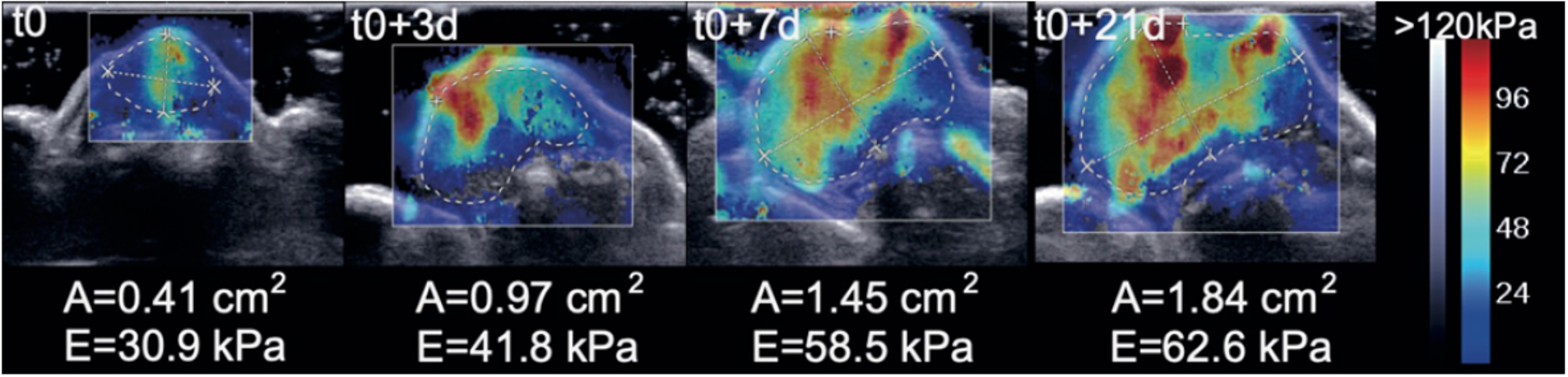
Representative colored stiffness maps in the transverse plan of Mesenchymal and Non-Mesenchymal PDX tumor growth over time. Colored map represents the Young’s modulus value. Mieulet, V., Scientific Reports (2021)
A fundamental question arise here: Once the cancer cell leaves their hometown, would they immediately forget their memories from there? Or is there any chance that the cells remembers the past mechanical microenvironments and be affected by it during the journey? To answer this question, we designed our experiment in consideration of two forementioned facts of ovarian cancer. This cancer model should experience either of soft or stiff substrate first, followed by a phase where they are completely isolated from solid substate and exist only as floating clusters.
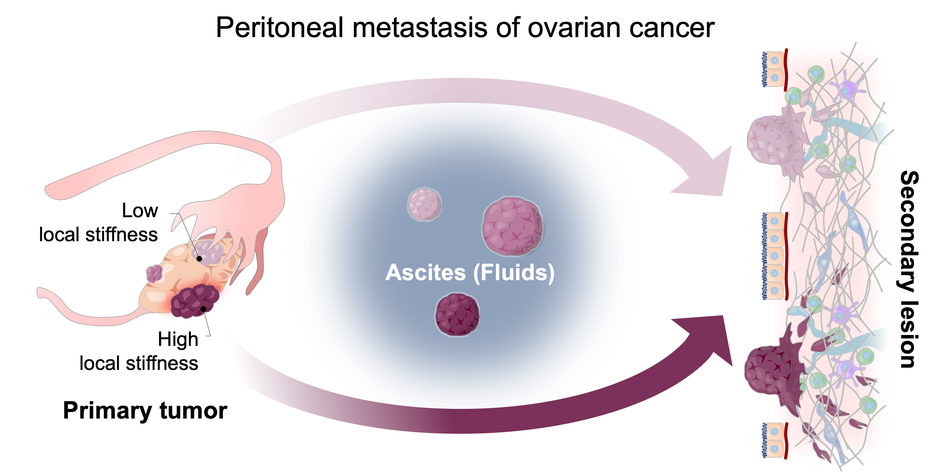
Peritoneal metastasis of ovarian cancer. Ovarian cancer cells exfoliate into peritoneal fluid forming multicellular spheroids, and float around until they reach a secondary lesion.
Briefly, we ‘educated’ the ovarian cancer cell on three different levels of stiffness, created hanging drop spheroids that resembles the in vivo malignant spheroids, and the finally tested whether the spheroids with different history show different invasiveness.
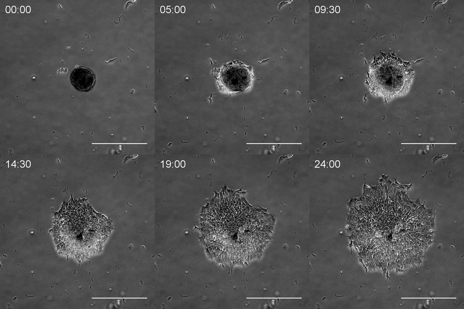
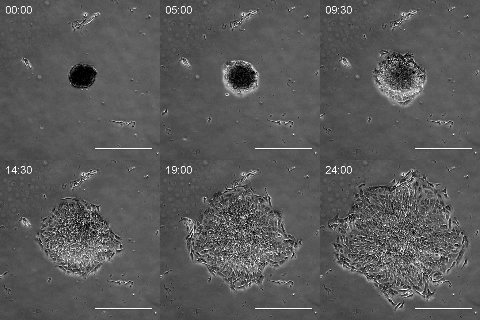
Spheroid invading on 3kPa PA gels. Spheroids made with low stiffness-primed cells (left), and high stiffness-primed cells (right). High stiffness-primed one spread faster.
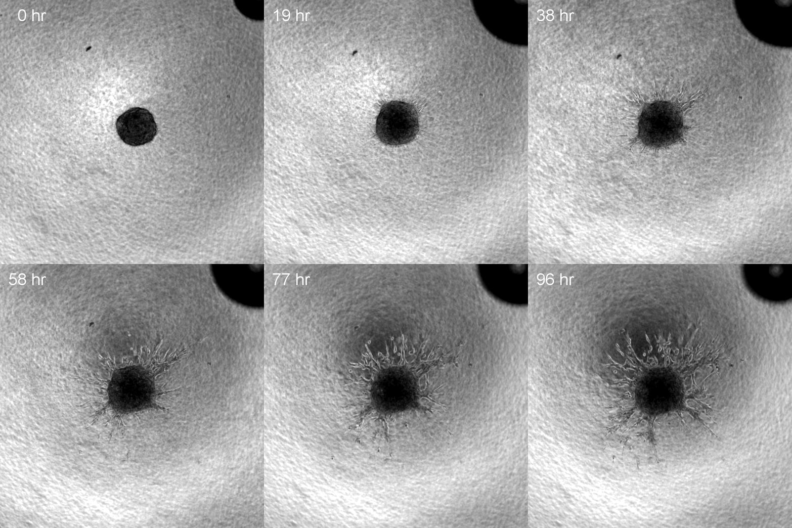
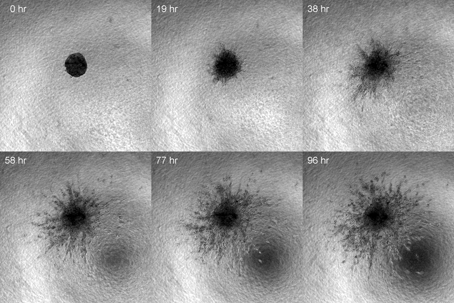
Spheroid invading into 3D collagen gel. Spheroids made with low stiffness-primed cells (left), and high stiffness-primed cells (right). High stiffness-primed one invade faster.